 By Emma Meyers
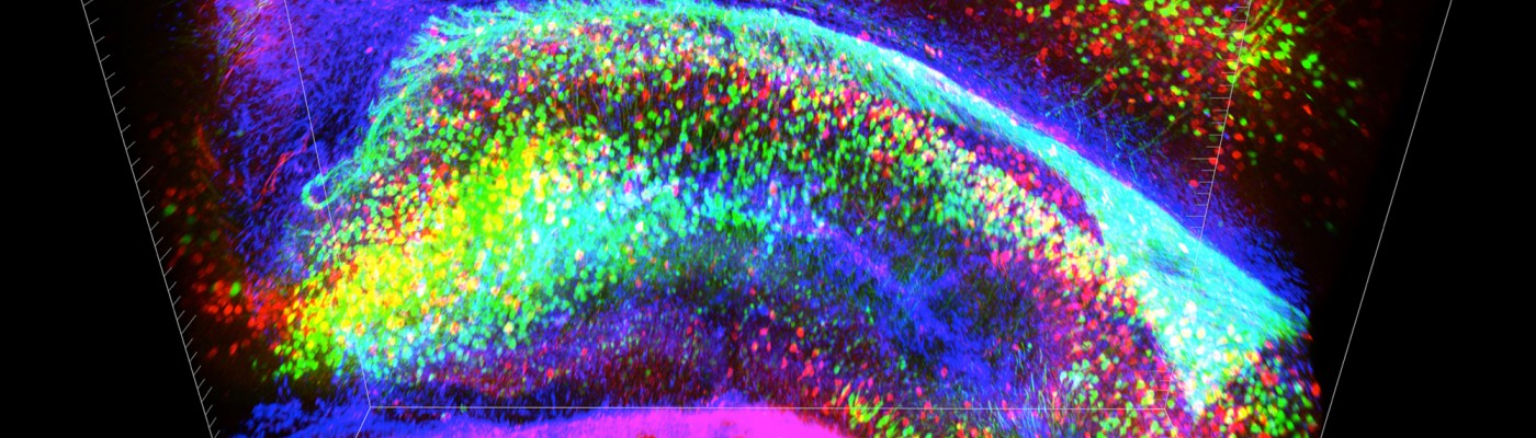
Ever since the Obama Administration announced its backing of a new $100 million initiative to map the connections of the brain’s 85 to 100 billion neurons, skeptics have questioned whether neuroscientists have the tools available to achieve the massive feat. Now, researchers at Stanford University are reporting in the journal Nature a new technology that renders a whole mouse brain transparent, with neural connections lit up by fluorescent antibodies to create a psychedelic 3D neuronal map. The process, developed by a team working under the direction of neuroscientist and psychiatrist Karl Deisseroth, is called CLARITY. It promises a new level of understanding of neural connectivity in a whole-brain context and can potentially be applied to make great strides in uncovering the mechanisms of still-mysterious mental disorders like schizophrenia and autism. Normally, brain tissue is opaque. Tightly packed fat molecules that make up the lipid bilayer encapsulating each cell act as walls that are impenetrable to molecular probes and scatter light, making imaging a challenge. The makers of CLARITY aimed to circumvent this problem by removing the troublesome lipids while still retaining the network’s structural integrity and important biomolecules like proteins. The transparent brain is created by infusing the tissue with hydrogel monomers, or hydrophilic molecules that interact with proteins and nucleic acids, but not lipids. When the hydrogel-tissue hybrid is heated, the monomers link together to form a stable three-dimensional network, locking the non-lipid components of the cells in place. Then, the loose fats are washed away with an ionic detergent and electric current, leaving a life-size, biochemically intact, and entirely clear brain. The standard methods researchers use to examine neurons involve cutting brains in extremely thin slices so that the individual cells can be seen through a microscope, separating cells from their neighbors and making piecing together each and every connection a painstaking and error-prone task. CLARITY is, therefore, a vast improvement when it comes to understanding connectivity in context. The transparency and mesh-like consistency of the hydrogel network make it the ideal antidote to the opacity of membrane lipids. The pores allow for deeper penetration of labeled antibodies than could be achieved when solid membranes were intact, and translucency provides for better visualization of deep structures under microscopes. These characteristics lend CLARITY to the study of proteins and other function-associated gene products, a valuable tool for researchers looking to identify the structural aspects of the active circuits they modulate in live animals. And, boding well for its practicality as a research tool, clarified brains are reusable; strong enough to endure the removal of injected probes, multiple proteins can be traced in the same brain. With all its promises, though, CLARITY is not without its limitations. For one, the process has only been implemented in full on mouse brains. While small portions of human brains have been successfully clarified, more work remains to be done before entire human specimens, much larger and more complex than mouse brains, can be made transparent. Additionally, as the authors of the journal article point out, CLARITY is not a replacement for microscopy. While it is invaluable for garnering a broad understanding of three-dimensional connectivity, it is a tool intended to be used in conjunction with standard practices of electron microscopy for high-resolution observation of the details of the synapses. Still, CLARITY holds a great deal of promise for neuroscience and other areas of molecular biology. As lead author Dr. Kwanghun Chung tells the New York Times, tissues other than the brain – the liver, heart, and lung, for example – can be clarified as well. With further refinement, CLARITY holds the key to a more complex understanding of the molecular intricacies of the human body than ever thought possible. For a video of Dr. Deisseroth explaining CLARITY, see Stanford School of Medicine’s website here.
0 Comments
Leave a Reply. |