|
By Tiago Palmisano
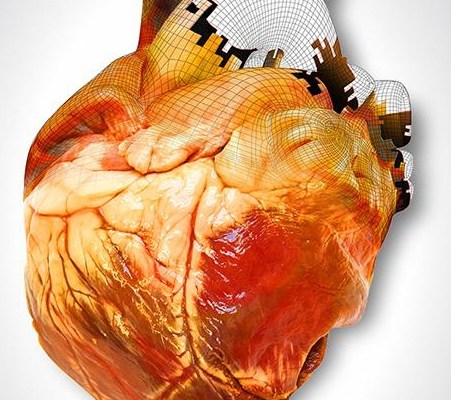
Integration of biological material with the technology of 3-D printing is beginning to redefine the limits of medical treatments and patient care, and will eventually change the ways that doctors treat patients. The last two decades have seen an explosive rise in the applications of 3-D printing–the process of making a three-dimensional object using a virtual design. 3-D printers use CAD (Computer Aided Design) files to deconstruct a product into thousands of successive 2-D layers, fabricating an object one layer at a time. Although the technology originated as a small manufacturing niche in 1986, it has grown substantially and is now commonly used to produce low-volume objects such as toys, wristwatches, and airplane parts. The majority of 3-D printers use some type of metal alloy, plastic, or powder to form each 2-D layer. Typically, a laser or heated nozzle is programmed to selectively harden the liquid or powder material into the desired shape out of the initially homogeneous substance. Recently however, more sophisticated printers and software have facilitated the use of increasingly complex materials such as biological cells. These advancements have ushered in a new age of “bioprinting” which is already transforming traditional medical care. Tissues fabricated with 3-D bioprinters, such as heart, skin and cartilage structures, are currently used as transplant components. In the past, fractured jaw and hip bones have been reconstructed using traditional material, such as titanium, but bioprinting is different. A biological 3-D printer generates actual tissue; the printed materials don’t mimic body parts, like metal, but rather are realcells, identical to the ones found in a human body. Scientists at Princeton University 3-D printed a functioning human ear using skeletal and cartilage cells.The first step to 3-D printing tissues is obtaining the digital file needed for the printing software. This is achieved with techniques such as computed tomography (CT) and magnetic resonance imaging (MRI) that gather extensive information of the tissue’s complex three-dimensional biological framework. Next, the 3-D printing software uses this data to carefully arrange living cells into the 2-D layers that form the intended shape, resulting in biologically compatible living tissue. In this case, “biologically compatible” means that the tissue does not induce a negative, inflammatory response in the body, making it ideal transplant material. A crucial part of the process is the selection of cells. Usually, bioprinters are loaded with stem cells–undifferentiated cells that have not yet been become specialized (an example of a specialized cell would be a muscle or blood cell). Because of this, stem cells can develop into the specific role required for the desired tissue. The chosen cells are also normally paired with a biologically compatible polymer for structural support. Although 3-D printing has successfully produced certain tissues, the ability to fabricate entire organs is still a vision of the future. Organs are extremely sophisticated structures with dozens of distinct cell types and functions, and just putting the right cells in the right shape usually isn’t enough to make a working organ. An organ must be able to deal with the intricate signals of the body and the intense stresses of the environment, such as heat or pressure. Despite this, the leap to full-fledged body parts such as organs may not be too far off. The amount of scientific literature referencing bioprinting has tripled in recent years, and research spending has increased significantly. Even if 3-D printed organs aren’t used in human transplants for many decades, other invaluable applications exist. Testing new drugs on fabricated organs could reduce the need for animal testing, and allowing medical students to train for surgeries using printed body parts would provide better preparation and lower patient risk. Additionally, the technology being developed for 3-D bioprinting may lead to other medical innovations. For example, labs at the University of Wollongong in Australia have created a handheld “bio pen”—a surgical tool that can deposit stem cells directly to damaged cartilage and bone. This provides doctors with a precise, efficient way to reconstruct certain tissues without the need to use cellular materials from other parts of the patient’s body. Indeed, the possible applications of 3-D bioprinting seem almost limitless. Quickly made and specific transplant material will vastly change how doctors heal and save lives. Though 3-D printed organs are not yet a reality, we may see them in the not-so-distant future. The need certainly exists, as over 500 people die waiting for transplants every month. Additional funding and research have facilitated great strides in the field of bioprinting. Such advancements intimate that a revolution in medicine is imminent, and given the improvements it may provide in patient care, it can’t come soon enough.
0 Comments
By Julia Zeh
When you put on sunscreen at the beach and then jump in the water, have you ever seen what looks like an oil slick appear in the water behind you? It turns out that sunscreen is not really all that waterproof, and it tends to come off when you go into the water. It’s not something that too many people notice or really begin to feel concerned about, but it just so happens that this small, sunscreen-induced oil slick actually has a negative impact on ocean life. Furthermore, components of sunscreen are found in noticeable quantities in areas around coral reefs. It may sound odd, but sunscreen can cause coral bleaching. The lotion you put on before you go swimming causes the loss of pigment and the eventual death of valuable marine organisms. Coral is very important because it is the foundation of the marine ecosystem; the lives of marine organisms depend on coral. Coral reefs are the home of rich biodiversity, and when something happens that impacts coral, the entire ecosystem that is based in that reef is impacted as well. We must protect our reefs because they are so important to many animals in the marine environment as well as to humans. Coral is also very sensitive, and a slight change in the conditions of the water in which it is living can drastically impact the coral. Coral bleaching, which more often than not leads to death of the coral, can be induced by changes in the water such as temperature, salinity, or pH. Bleaching occurs when coral’s symbiotic algae, zooxanthellae, is expelled. Zooxanthellae is a photosynthetic algal species that lives on the coral, providing it with food and giving it its color. When something goes wrong and causes stress to the coral, the coral’s response is to expel the zooxanthellae in an attempt to regain its health. When this occurs, the coral loses its pigment and it turns white, hence the name coral bleaching. After a coral bleaches, it generally will not return to good health because of the loss of its energy source, the zooxanthellae. Dr. Roberto Danovaro, a researcher in the Department of Marine Science at Polytechnic University of the Marche, Ancona, Italy, recently discovered that sunscreen has this impact on coral, causing bleaching and eventually death because of the effect of the chemicals that sunscreen contains. UV filters, a component in sunscreens and other cosmetics used on the skin, can contribute to coral bleaching. These compounds promote viral infections in the zooxanthellae, causing the coral to expel its symbiotic algae. After the algae is expelled, the coral will bleach and eventually die. In recent years, people have become increasingly obsessed with skincare items like sunscreen and cosmetics in an attempt to prevent skin cancer along with other forms of skin disorders. Along with this increased interest, however, come detrimental effects on the environment, a neglected but important relationship that is worthy of consideration. Although knowledge of thermal stress and rising ocean temperatures and their effects on coral is becoming more widely known, other sources of bleaching such as sunscreen are not as commonly understood. When people go for a swim at the beach, they’re not usually thinking about sunscreens or other cosmetics that might come off in the water, or how these products may impact the marine life around them. Yes, it’s nice to not get sunburned, but please be aware of what you’re putting on before you go in the water and the valuable marine life that may be affected. Author Alexander Bernstein
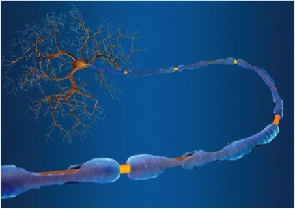
Editor Arianna Winchester Simply described as an autoimmune disease in which the body’s own defense mechanisms attack and degrade the protective myelin sheaths that cover axons in the nervous system, Multiple Sclerosis (MS) is estimated to effect some 250,000 to 350,000 people in the United States alone. The debilitating symptoms of the disorder include numbness and weakness in the limbs, loss of vision, body tremors, dizziness, finally building up to complete nervous system failure and death. Currently, despite how many people are affected by MS and a recent surge in donations towards MS research, there exists nothing even remotely close to a cure. At present, after a diagnosis is given to a patient, the only thing medical professionals can offer is symptom management. Further, there is still no complete understanding of what causes the disease. The only current medical knowledge claims that the disease is marked by an autoimmune response damaging the body’s own nervous system. Recent research has been seeking to reach a better understanding of what causes the disease. A presentation at the 2014 MS Boston Meeting suggested an interesting link between MS and the gut. It has previously been established that as much as 80 percent of the human immune system is at work in the gut, all as part of a complicated symbiotic relationship in which bacteria, fungi, and various single celled organisms contribute to the overall microbiome in the human body. A shift away from this delicate balance can cause a bevy of disorders like rheumatoid arthritis or diabetes. Regarding this connection between MS and the microbiome, a new study from the Brigham and Women’s Hospital is suggesting that single celled organism known as methanobrevibacteriaceae, present primarily in the gut, may play a role in the development of MS. Previous links between the MS and the microbiome have been found in US and Japanese studies. These studies have quantitatively demonstrated stark contrasts between the microbiomes of MS patients and the microbiomes of their healthy counterparts. Now, the recent study from the Brigham and Women’s Hospital reports that their baceriaceae, which is associated with activation of the immune system, is significantly augmented in the gut of MS patients. One exciting consequence of this work is that its conclusions may enable us to affect, or even drastically lower the onset of nerve diseases such as MS by modifying and establishing diets. Sushrut Jangi, Beth Israel Deaconess Medical Center physician and co-author of the study, emphasizes the potential importance of external factors on the microbiome and thereby autoimmune diseases, saying, “People who emigrate from non-Western countries, including India, where MS rates are low, consequently develop a high risk of disease in the U.S. One idea to explain this is that the biome may shift from an Indian biome to an American biome.” Further support for this connection arises from the effectiveness of the MS drug glatiramer acetate. In MS patients, the drug has proven to have the capacity to suppress excessive immune activity, thereby alleviating the effects of the disorder, though medical professionals do not know exactly how. A coalition of US research centers known as the MS Microbiome Consortium tested the drug in relation to the microbiome to try to elucidate a connection between the two body systems. After administering the MS drug, they saw a distinct change in gut flora populations. Although the role of inherent genetic differences in immune system function cannot be ruled out, the connection that has been demonstrated between MS and the microbiome holds great promise to eventually lead to external stimuli guidelines for how to minimize onset of the disease. |
Categories
All
Archives
April 2024
|


