 By Anna Christou
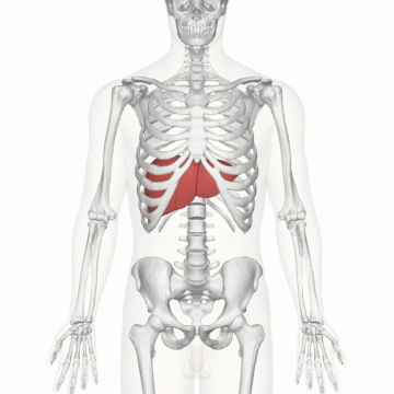
Physicians and scientists have been working to develop cancer therapies that are more personalized and that also minimize damage to healthy tissue. For example, researchers have recently developed strategies that either target the genetic mutation that causes the cancer or that harness the immune system’s natural killing ability in order to destroy tumor cells. Notably, embolization therapy, which consists of blocking blood flow to the tumor, has emerged as a novel treatment that is particularly useful for combating liver cancer. According to the American Cancer Society, the incidence of liver cancer, as well as its death rate, has increased dramatically in recent years. Specifically, it was projected that 42,200 new cases would be diagnosed in the United States in 2018 and that 30,200 people would die from this cancer. Liver cancer is especially difficult to treat with surgery because there are many blood vessels and bile ducts in the liver that make it risky. Therefore, embolization therapy—a minimally-invasive procedure performed by interventional radiologists to block blood flow to the tumor—offers a promising alternative to surgery. By preventing blood from reaching the tumor, embolization prevents the tumor from receiving the oxygen and nutrients that are necessary for its survival and growth. In addition to targeting the tumor cells more effectively than surgery, embolization therapy is also less invasive and less harmful to the surrounding blood vessels and bile ducts in the liver. This therapy also has several other advantages, including faster recovery time, no need for general anesthesia, no scarring, and lower risk of infection. While radiation therapy—which uses high energy waves to kill large numbers of cancerous cells while largely preserving the organ in question—is still an alternative to surgery, radiation damages the surrounding tissue, once again making embolization the best option. In order to block blood flow to the tumor, interventional radiologists, the physicians who perform embolization procedures, inject beads into the bloodstream that travel to the site of the cancer. The beads, which can be made up of a variety of materials such as gelatin, then physically block the artery, stopping the blood flow to the tumor. A challenge to this therapy is making sure to only block blood flow to the tumor, while leaving the blood supplied to healthy tissue intact. However, the anatomy of the liver circumvents this challenge and makes this therapy particularly advantageous for treating liver cancer. The liver is unique in that it is connected to two sources of blood. First, like other organs, the liver receives oxygenated blood from the heart through the hepatic artery. Second, the liver also receives blood directly from the digestive system through the portal vein so that it can process toxins that were ingested and passed through the digestive system. In this case, the blood supply to a certain liver cell depends on whether the cell is normal or cancerous; healthy cells receive blood from the portal vein whereas cancerous cells receive blood from the hepatic artery. As a result, in embolization therapy, only the blood supply from the hepatic artery is blocked, thereby reducing the blood—and consequently the oxygen and nutrients necessary for survival—of cancer cells, while leaving healthy cells intact. In addition to blocking blood flow to the tumor, embolization beads that contain added chemotherapy agents can further block and kill tumor cells through a technique called chemoembolization. By interfering with cell division, chemotherapy not only halts the growth of cancer cells but also stresses them to induce cell death. In particular, the chemotherapy agent doxorubicin, or adriamycin, is most often used along with embolization. This drug blocks topoisomerase 2, which is an enzyme that ensures that the two daughter cells resulting from cell division each contain the original DNA of the parent cell. As a result, by preventing DNA from being replicated, cell division is disrupted, halting the growth of the tumor. Delivering chemotherapy agents to the cancer cells through embolization targets the tumor cells more accurately to minimize the side effects that usually accompany chemotherapy, such as hair loss and nausea. Recently, chemoembolization has been taken to a new level with the addition of radiopaque beads, which allow for more precise and targeted treatments. A radiopaque substance blocks radiation, rather than letting it pass through, so it can be seen on x-rays. For example, bone, unlike air or fat, is radiopaque, which is why bone is visible as a white structure on an x-ray. Scientists create these beads by adding a radiopaque molecule to a gel such that the resultant bead can then block radiation. As a result, when these radiopaque beads, which could also contain chemotherapy as previously mentioned, are injected into the bloodstream, interventional radiologists can track the location of the bead through fluoroscopy, a type of x-ray that relays real-time moving images of the body. Therefore, rather than just deliver the chemotherapy-filled beads to the patient without knowing whether they are properly placed or not, doctors can instead view the position of radiopaque beads with x-ray images. This is especially important since blood vessel size and position may vary between patients, so the placement of the beads may not be the same for all people. As a result, because physicians can gain an inside look into the body and track the location of the beads, they can ensure that the bead is properly positioned and adjust it, if necessary, to provide a more targeted treatment to the specific patient. The Federal Drug Administration approved the use of radiopaque chemotherapy-filled beads for embolization procedures in 2015, and since then, hospitals have been conducting clinical trials to fine-tune the procedure, such as to determine the optimal length and amount of chemotherapy. Embolization therapy is a novel treatment that has emerged to target cancer cells effectively and precisely, while also minimizing damage to the surrounding healthy cells. Hopefully, this development for liver cancer treatment will pave the way for similar advancements in other types of cancer treatment.
0 Comments
Leave a Reply. |